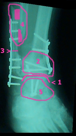
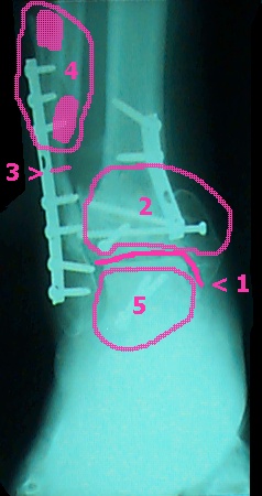
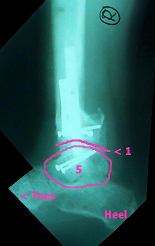
These numbers describe the areas in pink on the x-rays:
1 - the ankle joint
2 - bone graft area (this bone material was from my right hip)
3 - original break of the fibula
4 - The surgical re-break of the fibula to shorten my leg (about an
inch)
4 colored circles - note the filling in of bone material
5 - talus area. This bone is weak and may still cause problems
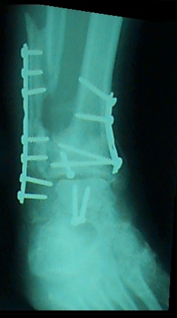
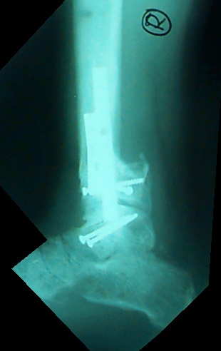
These are the first X-Rays that clearly show the hardware left in
my leg. It looks to be:
One plate with seven screws (holding the original break of the fibula
together)
One plate with three screws (bone graft area on the tibia)
Four individual pins / screws (two in the talus and two in the tibia
[as best I can tell])
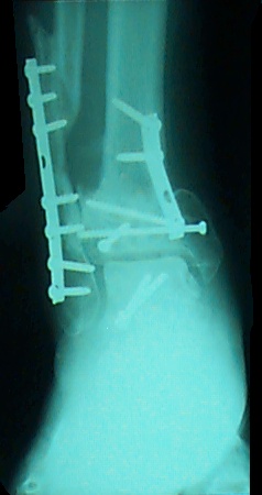
The top two rows of pictures are looking direct at the foot / ankle
(row 2 has just a slightly different angle). The bottom row shows
the inside of my right foot.
|
|
|
|
|
|